- Arthropoda (Arthropods)
- Mantodea (Mantids)
-
Insecta

The eyes of the scorpion
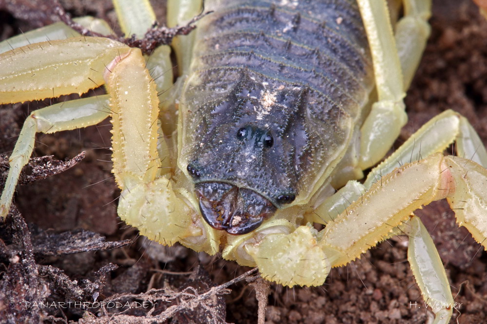
The two median und six lateral eyes of Hottentotta buchariensis
The main visual sensory organs of a scorpion are its two median and several lateral eyes, alongside the extraocular light sense in its metasoma (Zwicky, 1968). The former are located on either side of the median plane of the carapace usually in the vicinity of the mid line, but much more anterior in case of some species (Polis, 1990, p.30). The number of lateral eyes varies from zero to ten, they are situated on the anterolateral margin of the carapace, usually in a paired manner (Polis, 1990, p.30). There are however some relatively few species, that have lost their eyes alltogether in the course of adaptation to dark environments.

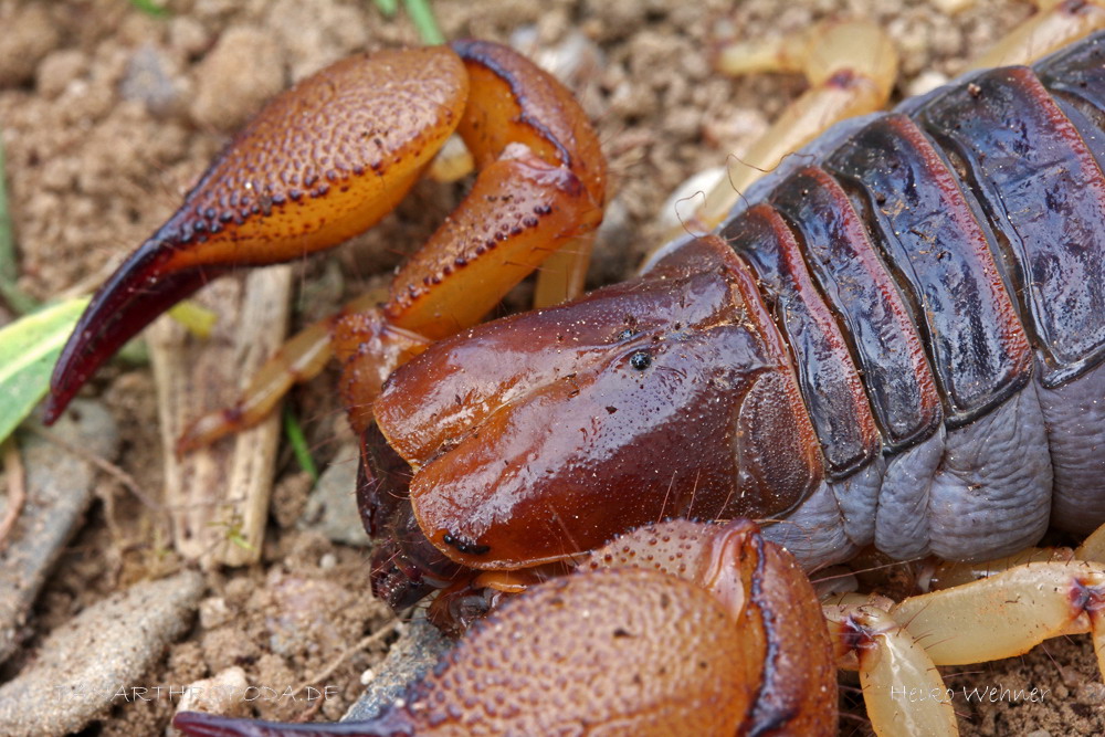
A totally eyeless species of scorpion: Belisarius xambeui
The median eyes consist of a well visible lense which lies on top of a vitreous body, divided only by a preretinal membrane (Machan, 1967). Behind this vitreous body lie the rod-shaped photoreceptor cells (Retinula) of the retina, that gather the light signals.
On the distal region (close to the vitreous body) these retinula possess rhabdomeres, a seam of cell protrusions, which link between four and six retinula into one retinula complex - the individual complexes bear no such rhabdomere connections (Schliwa and Fleissner, 1980). Inside of the retinula cells one finds a multitude of pigment granules, which migrate through the cell depending on several conditions, located proximally inside the cell lies the nucleus, the optical nerve attaches on the outer proximal side. The retinula cells are sparcely surrounded by pigment cells and are in total enfolded, thus completing the retina, by the postretinal layer (Machan, 1967).
The lateral eyes show significant differences to this composition. They lack the vitreous body alltogether and all retinula cells are connected by rhabdomeres into one single retinula complex (Schliwa and Fleissner, 1980). Furthermore, the postretinal layer of the lateral eyes bears pigment cells as well (Machan, 1967). Lastly the lateral eyes have no direct connection with each other, whereas the median eyes are directly connected by an additional nerve (Machan, 1967).
The name of its genus already suggests the anteriorely skewed position of the median eyes: Opistophthalmus glabrifrons
The lack of the vitreous body and the high interconnectivity of the retinula of the lateral eyes cause a reduced contrast (physically by the lack of the vitreous body, neurologically by the crossfiring of neighbouring retinula cells) and thus suggest that the lateral and median eyes have different biological uses (Schliwa and Fleissner, 1980). Furthermore, the optical structure of the lateral eyes only allows for low spatial discrimination (Carricaburu, 1968).
One possible explanation of these findings is that the lateral eyes are used mainly as light sensors and as Zeitgebers for the circadian rhythm (internal clock) (Fleissner, 1977a). This is indicated by physiological experiments and the fact, that due to the high interconnectivity of the retinula cells the lateral eyes are much more sensitive towards changes in luminance even at very low intensities (Fleissner, 1977b).
In addition, it has been shown that the pigment granules inside the retinula migrate in accordance to the circadian rhythm - they are located on the distal end near the vitreous body during the day phase and near the nucleus during the night phase (Schliwa and Fleissner, 1980). This migration is highly reduced if the scorpion is exposed to lasting darkness in case of the lateral eyes but remains virtually unchanged in case of the median eyes (Fleissner, 1974). Thus the lateral eyes react much more adaptively towards changes in light stimuli than the median eyes.
This fact is reflected by the absolute sensetivities of the median and lateral eyes. The median eyes of tested specimen showed a absolute sensitivity of 2,7e-3 cd/m² during the day phase and no significant change of sensetivity during the night phase, whereas the sensitivity of the lateral eyes increased from 5,5e-3 cd/m² during the day phase to 0,86e-3 cd/m² and in some cases even to 0,35e-3 cd/m² during the night phase - sensitivities that even surpass those of the human eye (Fleissner, 1977b).
All these insights allow for the assumption, that scorpions possess two different visual systems - one encompassing high contrast and high spatial discrimination (the median eyes) and one with high absolute sensitivity (the lateral eyes) (Schliwa and Fleissner, 1980).
References
Fleissner, G. (1974). Circadiane Adaptation und Schirmpigmentverlagerung in den Sehzellen der Medianaugen von Androctonus australis L. (Buthidae, Scorpiones). J. Comp. Physiol. 91, 399-416.
Fleissner, G. (1977a). Scorpion lateral eyes: extremely sensitive receptors of Zeitgeber stimuli. J. Comp. Physiol. 118, 101-108.
Fleissner, G. (1977b). The absolute sensitivity of the median and lateral eyes of the scorpion Androctonus australis L. (Buthidae, Scorpiones). J. Comp. Physiol. 118, 109-120.
Machan, L. (1967). Studies on the structure and electrophysiology of scorpion eyes. South African Journal of Science (Dec), 512-520.
Polis, G.A. (1990). The Biology of Scorpions. Stanford University Press, Standford, California.
Schliwa, M. und Fleissner, G. (1980). The Lateral Eyes of the Scorpion, Androctonus australis. Cell Tissue Res. 206, 95-114.
Zwicky, K.T. (1968). A light response in the tail of Urodacus, a scorpion. Life Sci. 7, 257-262.
F. Schmitz, authored 2012